Li Chengyu, a researcher at the Center for Excellence in Brain Science and Intelligent Technology of the Chinese Academy of Sciences, said that it should be said that this is a very important milestone in the entire field of neuronal injury and regeneration. It is of great significance for us to develop new intervention methods to treat brain injury in the future.
Maria Antonietta Tosches, assistant professor at Columbia University, commented that this paper proves that spatiotemporal omics technology with unprecedented spatial and single-cell resolution can open new perspectives on biological problems such as brain regeneration. 
Cover of Science. The Mexican axolotl, also known as the "hexagonal dinosaur", has a powerful regeneration ability.
The regeneration of cells and organs is a crucial topic for regenerative medicine. That means whether a paralyzed person can get back on his feet, a person with liver disease or heart failure can regain their health.
But can nerve cells, even damaged organs, or even the brain, be regenerated? How to achieve this?
Achieve a Grand Slam in half a year
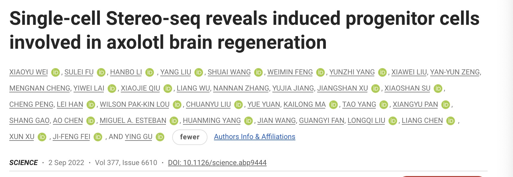
On September 2, the first spatiotemporal map of salamander brain regeneration, jointly completed by BGI, Guangdong Provincial People's Hospital, and Wuhan University, was published online in the top international academic journal Science in the form of back-to-back cover articles. ). This is also the world's first spatiotemporal map of brain regeneration.
Elly Tanaka, a senior scientist at the Institute of Molecular Pathology in Austria, said the research is important for those interested in nerve regeneration. Its findings show that during successful organ regeneration, not only neural stem cells respond to injury, but even surrounding neurons to create an important microenvironment. The findings provide inspiration for researchers who want to induce regeneration in other vertebrates, even humans.
So far, in just half a year, BGI's results related to spatiotemporal omics and single-cell technology have been published in the three top journals "Cell", "Nature" and "Science" for four consecutive times. Achieved a grand slam.
Five months ago, on April 13, Shenzhen BGI Life Sciences Research Institute published a paper online in the international academic journal "Nature". Based on single-cell library construction and sequencing analysis, it reported the whole body organ cell map of macaques. This is also the world's first non-human primate whole-cell map.
Four months ago, using the spatiotemporal omics technology Stereo-seq, on May 4, research teams including Shenzhen BGI Life Sciences Research Institute jointly released the world's first spatiotemporal maps of life. The paper on the spatiotemporal map of mouse embryonic development was published online in the journal Cell, and the related results on the spatiotemporal map of zebrafish, Drosophila, and Arabidopsis were published online in Developmental Cell, a sub-journal of Cell.
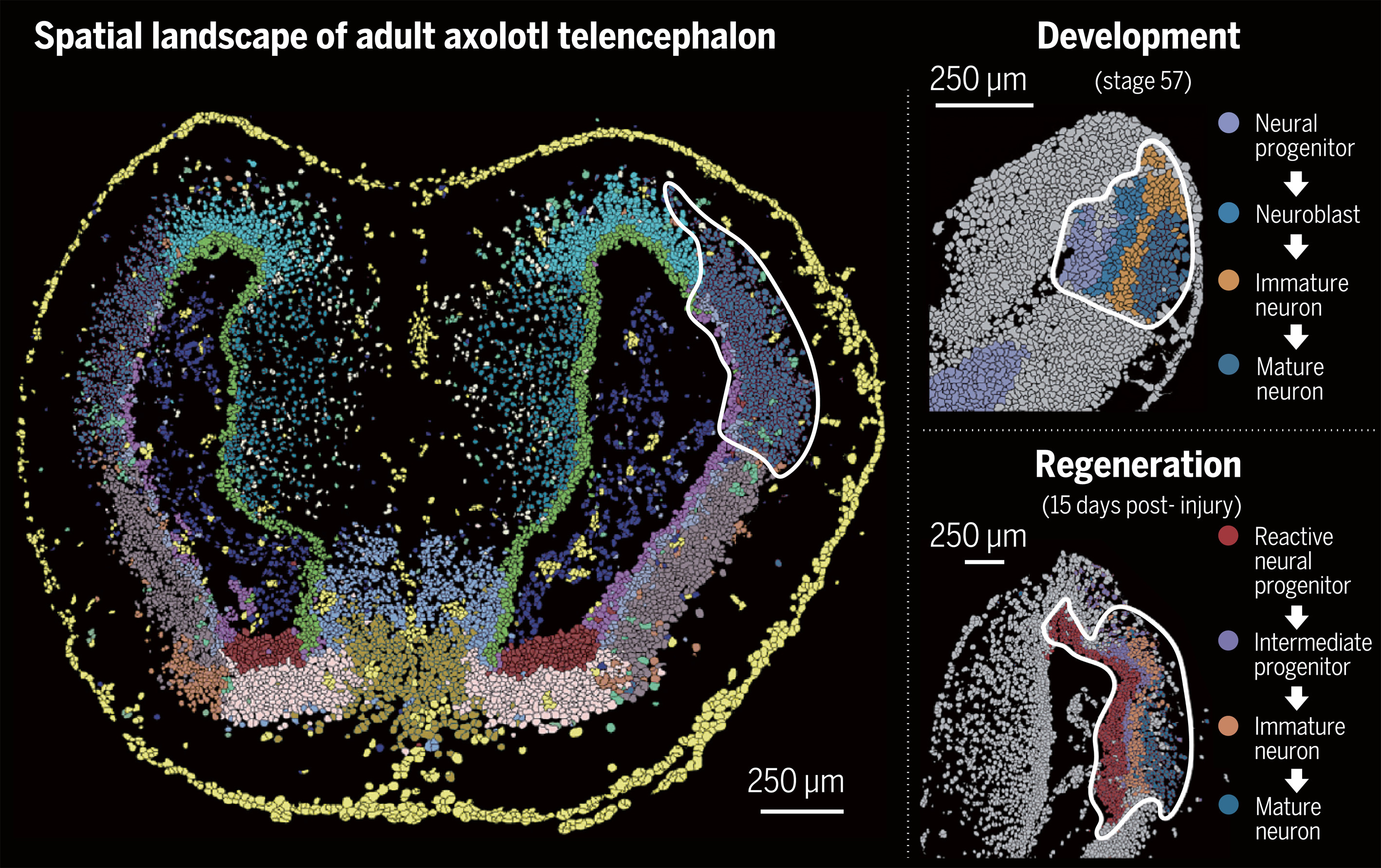
In the aforementioned paper published in Science, the research team systematically analyzed and compared the development and regeneration of salamander brains based on BGI's Stereo-seq technology, and found the key neural stem cells in the process of salamander brain regeneration. Subpopulations, depicting the process of remodeling damaged neurons by such stem cell subpopulations, provide new directions for regenerative medicine research and treatment of the nervous system.
Stereo-seq technology is called the "ultra-wide-angle 10-gigapixel life camera", which can simultaneously "photograph" the genetic information and spatial location of each cell in the tissue. The secret is in its chip.
The chip used in the Stereo-seq technology is a DNA nanosphere space capture chip with spatial position information and arrayed based on the DNBSEQ sequencing technology. The chip can achieve ultra-high precision and ultra-large field of view for molecular imaging of life, with a resolution of up to 500 nanometers. 
The world's first paper on brain regeneration spatiotemporal map was published online in the academic journal Science on September 2.
Tracing the secrets of brain regeneration in 'hexagonal dinosaurs': spatial trajectories of cell lineage changes
The research object of the aforementioned latest research paper is a salamander, the Mexican axolotl, also known as the "hexagonal dinosaur", with a unique and cute appearance.
The Mexican axolotl has powerful organ regeneration capabilities: not only can it regenerate organs such as limbs, tail, eyes, skin, and liver, but it can even regenerate the brain.
What can it teach people?
Dr. Gu Ying, the co-corresponding author of the paper and the Hangzhou BGI Institute of Life Sciences, said that the genetic coding sequence of salamanders is very similar to humans, and has a high similarity to mammalian brain structure. Therefore, studying the initiation mechanism of salamander brain regeneration and discovering the key genes may provide important guidance for the repair of human nervous system damage or degenerative diseases.
Compared with past microscopy and sequencing technologies, spatiotemporal omics technology can simultaneously observe cell morphology and tissue morphology, and can comprehensively detect gene transcriptomes at the molecular level, realizing the understanding of cell function from the perspective of high-precision structure. "Therefore, we used spatiotemporal omics technology to study the brain regeneration of the Mexican axolotl, and realized the in situ observation and analysis of the dynamic changes of the key stem cells that are really involved in the regeneration near the injury site in the regeneration of the brain injury." Article communication The author, Fei Jifeng, a professor at Guangdong Provincial People's Hospital, said.
Dr. Xiaoyu Wei, the first author of the paper and the Hangzhou BGI Institute for Life Sciences, said that this study analyzed the important cell types in the process of brain regeneration in salamanders at the spatiotemporal single-cell resolution, and tracked the space of cell lineage changes. Trajectories, mainly based on Stereo-seq technology, which achieves nanoscale subcellular resolution and the large size of salamander cells.
"The construction of a spatiotemporal cell map of salamander brain development and regeneration is of great significance for us to understand the important life process of brain regeneration, the brain structure of amphibians and the evolution of brain structure, and to find effective clinical treatment methods for us to promote human beings. The self-repair and regeneration of tissues and organs provides a new direction, and also provides valuable data resources for species evolution research." Xu Xun, the co-corresponding author of the paper and dean of the BGI Institute of Life Sciences, said, "In the future, we will also use Spatiotemporal multi-omics technology can explore the development and regeneration process of more organs and species, find the key regulatory mechanism in the regeneration process, and help the development of human regenerative medicine."
The research was in-depth cooperation between Shenzhen BGI Life Sciences Research Institute and Guangdong Provincial People's Hospital Fei Jifeng's team, in conjunction with South China Normal University, Wuhan University, School of Life Sciences, University of Chinese Academy of Sciences, Shenzhen Bay Laboratory, Whitehead Biomedical Research Institute, Denmark The University of Copenhagen and other units from China, the United States, and Denmark cooperated to complete the project.
In order to study the regeneration process of salamander brain injury, the research team performed mechanical injury surgery on the cortical area of salamander brain, and performed 7 time points of regeneration (2, 5, 10, 15, 20, 30 and 60 days after injury). ) brain samples for analysis. The spatiotemporal data showed that a new subset of neural stem cells emerged in the wound area early in the injury.
By comparison, the researchers found that the neuron formation process during the development and regeneration of the salamander brain is highly similar, suggesting that brain damage may induce the reverse transformation of the salamander neural stem cells, returning to the "young" stage and starting the regeneration process.
In addition, the research team constructed a spatiotemporal map of six important developmental periods of the salamander brain, showing the molecular characteristics of various neurons and the dynamic changes in spatial distribution, and found that the salamander brain began to specialize in spatial regions from adolescence of neural stem cell subtypes. 
Development and regeneration of the salamander telencephalon.
Unprecedented resolution of new technology brings new perspectives to help understand brain injury and regeneration
In response to this newly published research result, Zeng An, a researcher at the Center for Excellence in Molecular and Cell Science of the Chinese Academy of Sciences, who is mainly engaged in the mechanism research of tissue and organ regeneration, said that this is a very important work. First, it takes full advantage of the large field of view and high resolution of spatiotemporal omics, and develops corresponding algorithms to obtain cell lineages and spatiotemporal dynamic changes during the regeneration process of the full field of view. This technique has important reference significance for other tissue and organ regeneration or regeneration model research.
Zeng An said, secondly, the study also found that there are stress-induced neural progenitor cell types during the regeneration of the axolotl telencephalon. It sheds light on the source of cells during wound healing and tissue remodeling following extensive damage to the axolotl telencephalon. This has important implications for our understanding of brain injury and brain regeneration in higher organisms.
Li Chengyu, a researcher at the Center for Excellence in Brain Science and Intelligent Technology of the Chinese Academy of Sciences, said that it should be said that this is a very important milestone in the field of neuron damage regeneration. This work points out a very important direction for us to achieve brain damage repair in mammals, especially in primates in the future, and is of great significance for our future development of new intervention methods to treat brain damage.
Maria Antonietta Tosches, an assistant professor at Columbia University, commented that BGI has invented a new technology - spatiotemporal omics technology. This paper demonstrates that spatiotemporal omics techniques with unprecedented spatial and single-cell resolution can open new perspectives on biological problems such as brain regeneration. At the same time, this paper also confirms that this new technology can be used in any species and any organ, and can also lead to new discoveries in the biological fields of brain development, brain evolution or wider development, evolution, neuroscience, regeneration and other biological fields. .


