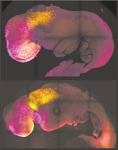
Comparison of naturally formed and synthetic embryos. Image source: "Nature" online edition
According to a paper published in the British "Nature" magazine on August 25, a joint team including the California Institute of Technology in the United States, the University of Cambridge in the United Kingdom and other institutions described the generation of a synthetic mouse embryo derived from stem cells. This embryonic model replicates the developmental stages of a natural mouse embryo from fertilization to day 8.5, including the formation of clear brain regions, a neural tube, and a beating heart-like structure. In addition, the model was able to replicate the gene knockout results observed in natural mouse embryos. This finding raises the possibility that such models could be used to understand the factors that regulate early stages of development without the use of laboratory animals.
Embryonic stem cells are able to form embryo-like structures in the laboratory. But these models cannot fully simulate all stages of development, for example, they cannot fully recapitulate a process called neurulation (the formation of the neural tube, which eventually differentiates into the brain and spinal cord).
The researchers assembled stem cell-derived mouse embryos in the laboratory this time, using a combination of embryonic stem cells, trophoblast stem cells and induced extraembryonic endoderm stem cells, all of which were derived from mice. The resulting ETiX embryoid body model was able to develop to post-neurogenesis, the equivalent of a natural mouse embryo at 8.5 days post-fertilization, and formed all brain regions, a neural tube, a beating heart, and an intestinal tube.
The research team mentioned that the model was able to achieve this result with self-organizing stem cell types without external signal modulation. In further experiments, the team successfully demonstrated an embryoid body model in which the Pax6 gene, which is involved in the development of the eye and other sensory organs, was knocked out, resulting in similar effects to natural Pax6 knockout mouse embryos.
The researchers concluded that ETiX embryoid bodies provide a useful physiological model of embryonic development, providing new opportunities to study developmental and disease mechanisms.
[Editor-in-chief's circle]
The current study, which is at the most advanced stage to date achieved in stem cell-derived models, has a key advance in generating capacity equivalent to the entire mouse brain, specifically duplication of the anterior brain region, which has been synthetic embryos The "crown" of the developmental world. The real significance of the method is twofold. One is to help researchers understand why some embryos fail and others develop smoothly. The other is that, if successful in human stem cells in the future, it can be used to guide the synthesis of organs. Repair and development to help save the lives of a large number of patients waiting for transplantation.
(Original title "Stem-cell-derived synthetic mouse embryos generate clear brain regions, neural tubes, and beating heart-like structures")


Comments